Comprehensive Dengue Clinical Reference Guide
As dengue remains one of the most critical public health and emergency medicine challenges in Bangladesh and across tropical regions, early bedside vigilance and rigorous hemodynamic monitoring are essential to saving lives. The Dhaka Medical College Internal Medicine Interest Group (DMC IMIG) has developed this consolidated clinical reference guide, translating international guidelines (WHO, SEARO, and National Guidelines) into a structured, actionable bedside framework for medical students, house officers, and clinical trainees.
This unified masterclass synthesizes the complete clinical trajectory: disease classification, bedside warning signs, plasma leakage pathophysiology, hematological monitoring, diagnostic testing windows, organ complications, and life-saving critical care protocols.
Section 1: The Clinical Spectrum of Dengue Infection
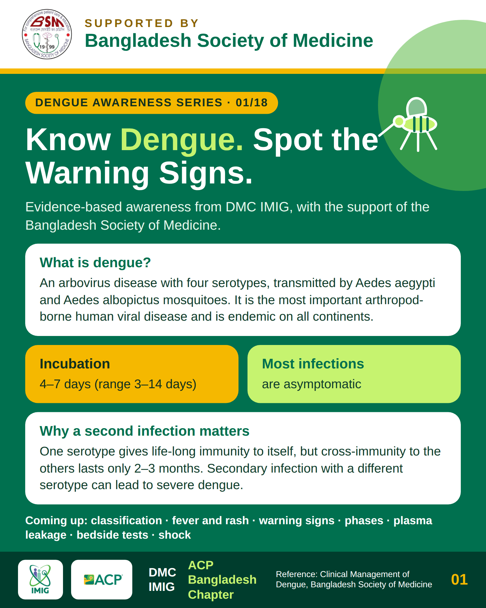
Dengue virus (DENV-1 through DENV-4, family Flaviviridae) infection causes a wide spectrum of illness, ranging from completely inapparent or asymptomatic infection to severe, life-threatening multi-organ failure.
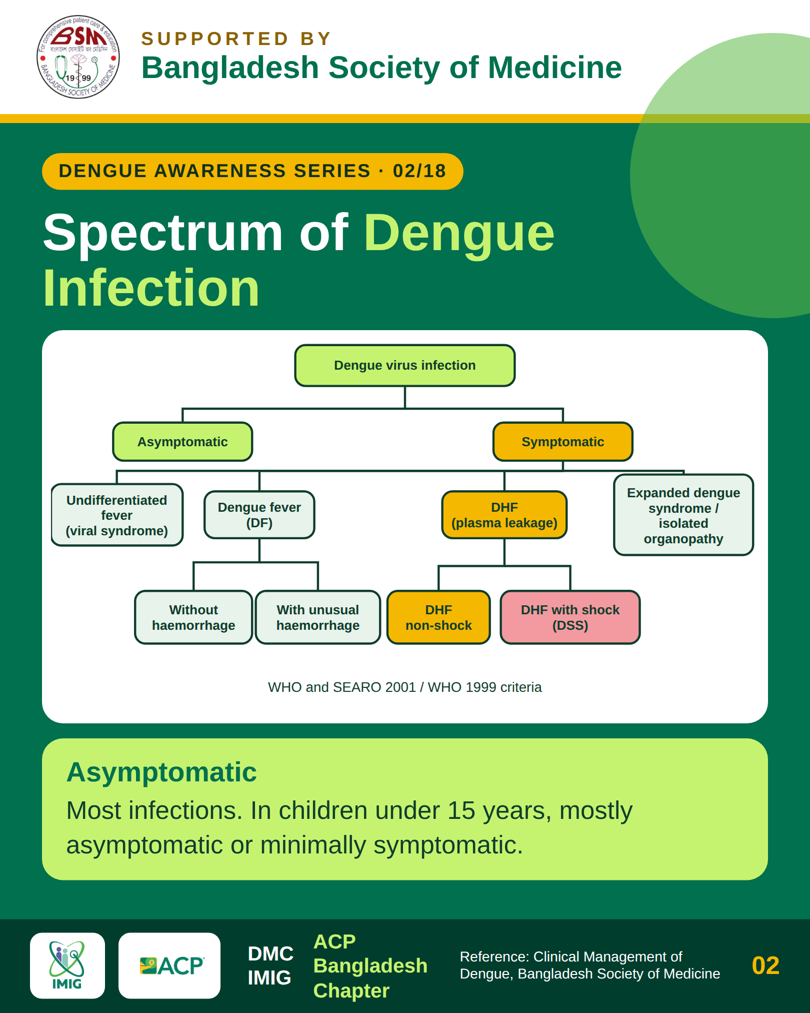
Clinically symptomatic patients are categorized into three major clinical presentations:
- Dengue Fever (DF): Severe acute febrile illness accompanied by headache, retro-orbital pain, myalgia, arthralgia, bone pain ("breakbone fever"), and macular or maculopapular rash, without significant plasma leakage.
- Dengue Haemorrhagic Fever (DHF): Characterized by four hallmark criteria: sustained fever, hemorrhagic tendency (positive tourniquet test or spontaneous bleeding), thrombocytopenia (platelet count ≤ 100,000/uL), and objective evidence of plasma leakage (hematocrit rise ≥ 20%, pleural effusion, or ascites).
- Grade I: Positive tourniquet test as the only hemorrhagic manifestation, alongside thrombocytopenia and plasma leakage.
- Grade II: Spontaneous bleeding (mucosal, cutaneous, or gastrointestinal), thrombocytopenia, and plasma leakage.
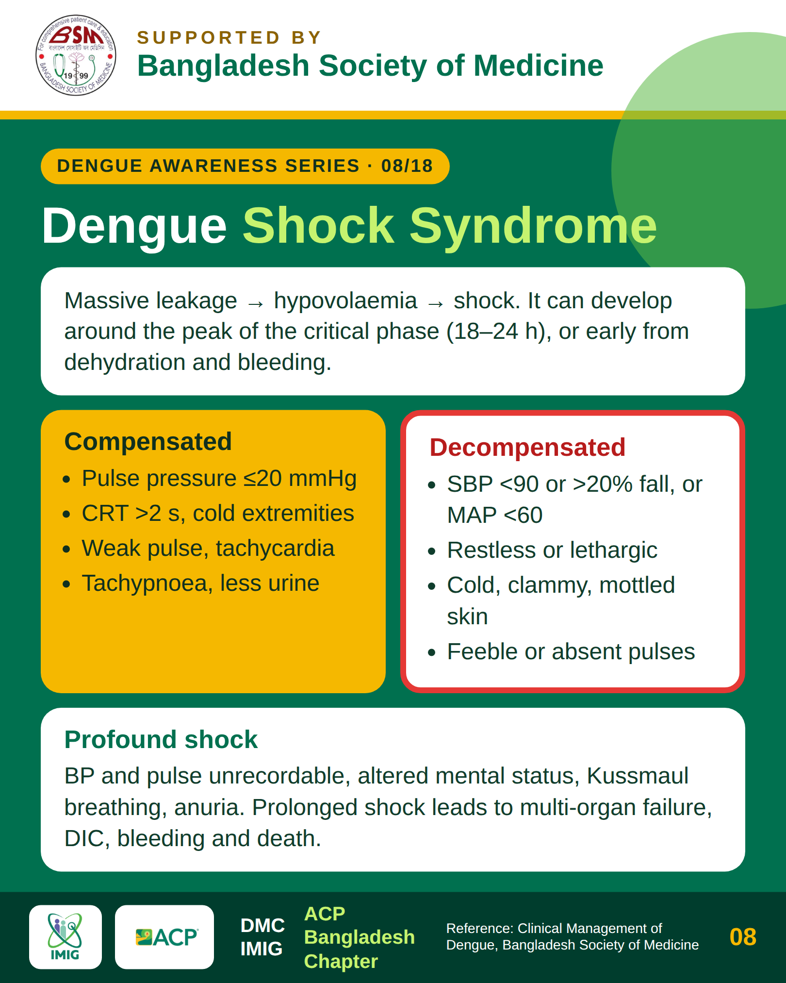
- Grade III (Compensated Shock): Circulatory failure characterized by a rapid, weak pulse and narrowing of pulse pressure (≤ 20 mmHg) or hypotension, cold clammy extremities, and restlessness.
- Grade IV (Decompensated Shock): Profound, unrecordable blood pressure and undetectable peripheral pulse.
- Expanded Dengue Syndrome (EDS): Atypical and severe clinical presentations involving profound single or multi-organ failure (hepatic failure, acute encephalitis, myocarditis, or renal shutdown) with or without typical plasma leakage.
Section 2: The Three Phases of Dengue Haemorrhagic Fever
Understanding the timeline of DHF is essential because the most dangerous phase occurs precisely when the fever breaks.
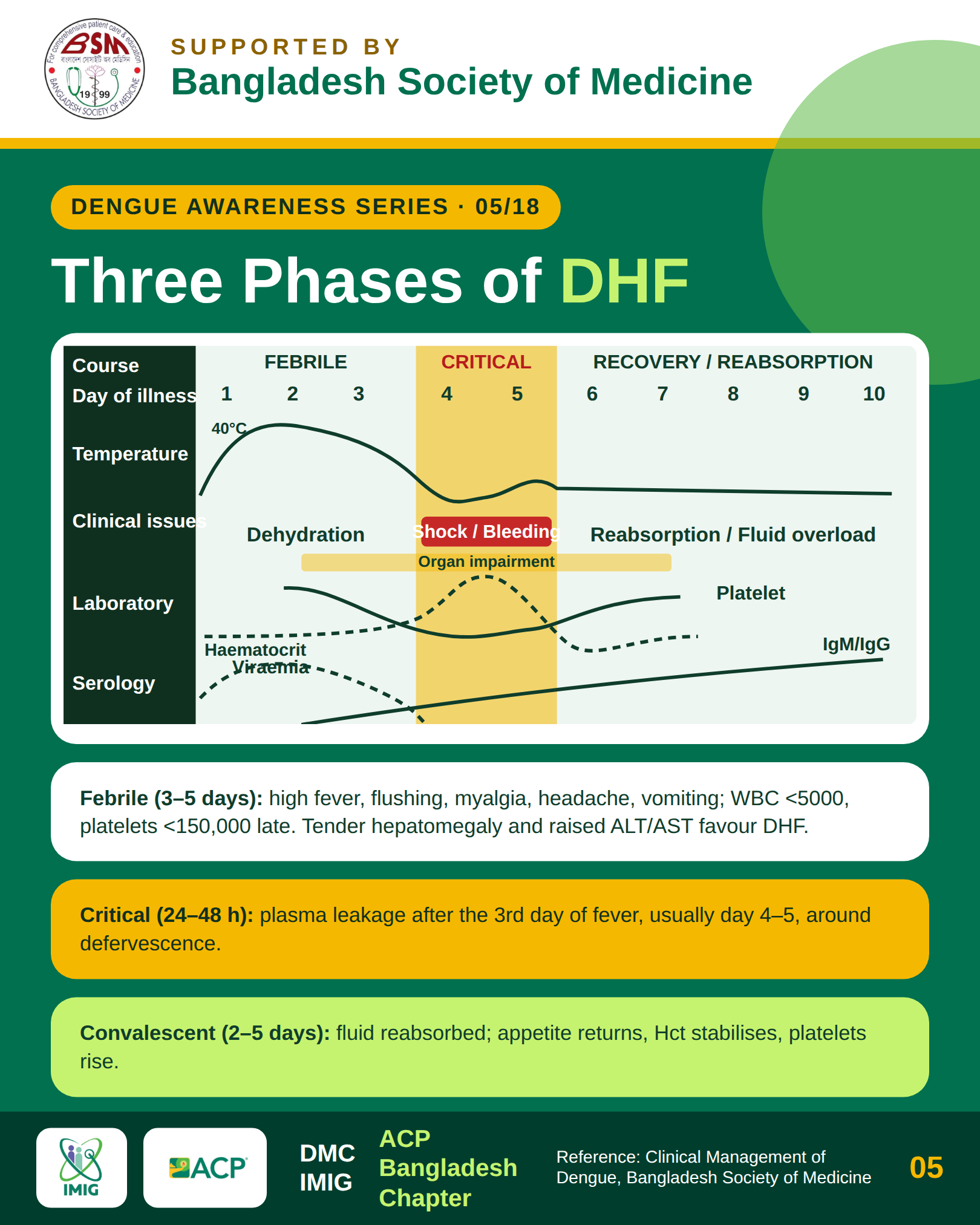
Phase 1: Febrile Phase (Days 1 to 3)
- High-grade, abrupt-onset continuous fever (often 39 to 40 deg C), accompanied by facial flushing, cutaneous erythema, retro-orbital headache, and widespread muscular tenderness.
- Mild hemorrhagic manifestations (petechiae, mucosal epistaxis) may appear.
- Leukopenia is typically the earliest hematological indicator, frequently dropping 24 to 48 hours prior to platelet reduction.
Phase 2: Critical Phase (Days 3 to 6, Lasting 24 to 48 Hours)
- Coincides with defervescence (temperature dropping to 37.5 to 38 deg C or below). Patients and inexperienced clinicians may mistake defervescence for clinical recovery.
- Significant plasma leakage into the pleural and peritoneal cavities occurs due to selective endothelial hyperpermeability.
- Hematocrit surges in direct proportion to the volume of intravascular fluid lost.
- If fluid losses are uncompensated, patients slide rapidly into compensated and then decompensated shock.
Phase 3: Convalescent Phase (Days 7 to 10)
- Spontaneous cessation of plasma leakage with active reabsorption of extravasated fluid back into the intravascular compartment.
- Appetite returns, gastrointestinal symptoms abate, and hemodynamic parameters stabilize.
- A characteristic convalescent confluent petechial rash ("islands of white in a sea of red") frequently emerges with intense pruritus.
- Clinical Caution: Continuing aggressive intravenous fluid therapy during this phase carries high risk of iatrogenic hypervolemia, acute pulmonary edema, and congestive cardiac failure.
Section 3: Bedside Warning Signs & Triage Triggers
The presence of any warning sign mandates urgent hospital admission, close hemodynamic monitoring, and initiation of intravenous fluid titration.
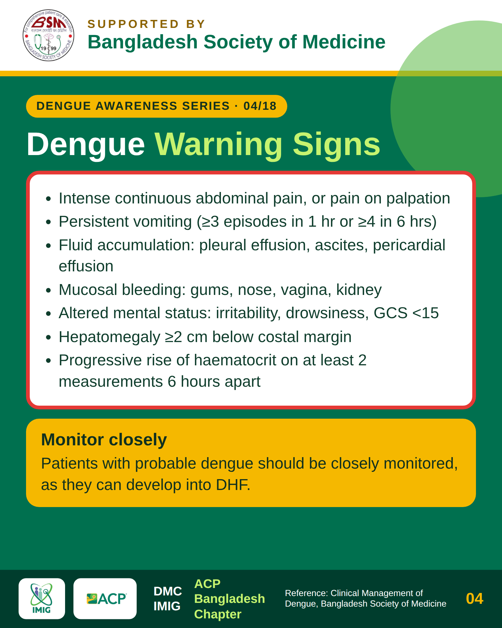
- Severe Abdominal Pain or Tenderness: Caused by thickening of the gallbladder wall, subcapsular hepatic swelling, or early peritoneal fluid collection.
- Persistent Vomiting: Defined as ≥ 3 episodes within 24 hours, preventing adequate oral rehydration.
- Clinical Fluid Accumulation: Detectable pleural effusion on chest radiography/ultrasonography or clinical ascites.
- Mucosal Bleeding: Spontaneous epistaxis, gingival bleeding, hematemesis, melena, or abnormal menorrhagia.
- Lethargy, Restlessness, or Altered Mental Status: Manifestations of cerebral hypoperfusion or early metabolic encephalopathy.
- Hepatomegaly (> 2 cm Below Costal Margin): Reflecting tender hepatic congestion.
- Progressive Hematocrit Rise with Rapidly Dropping Platelets: Laboratory evidence of accelerating plasma extravasation.
Section 4: Pathophysiology of Plasma Leakage & Shock Dynamics
Unlike septic shock or capillary leak syndromes caused by direct microbial endotoxins, plasma leakage in dengue is unique: it is transient, immunologically driven, and characterized by the absence of structural vascular endothelial necrosis.
During secondary infection with a heterologous DENV serotype, pre-existing non-neutralizing antibodies cross-react and bind to the newly infecting virus, facilitating viral uptake into Fc-receptor-bearing monocytes and macrophages (Antibody-Dependent Enhancement, ADE). This triggers an uncontrolled surge of pro-inflammatory mediators (TNF-alpha, IL-6, IL-8, IL-10) and secreted viral non-structural protein 1 (NS1), which disrupts the endothelial glycocalyx layer.
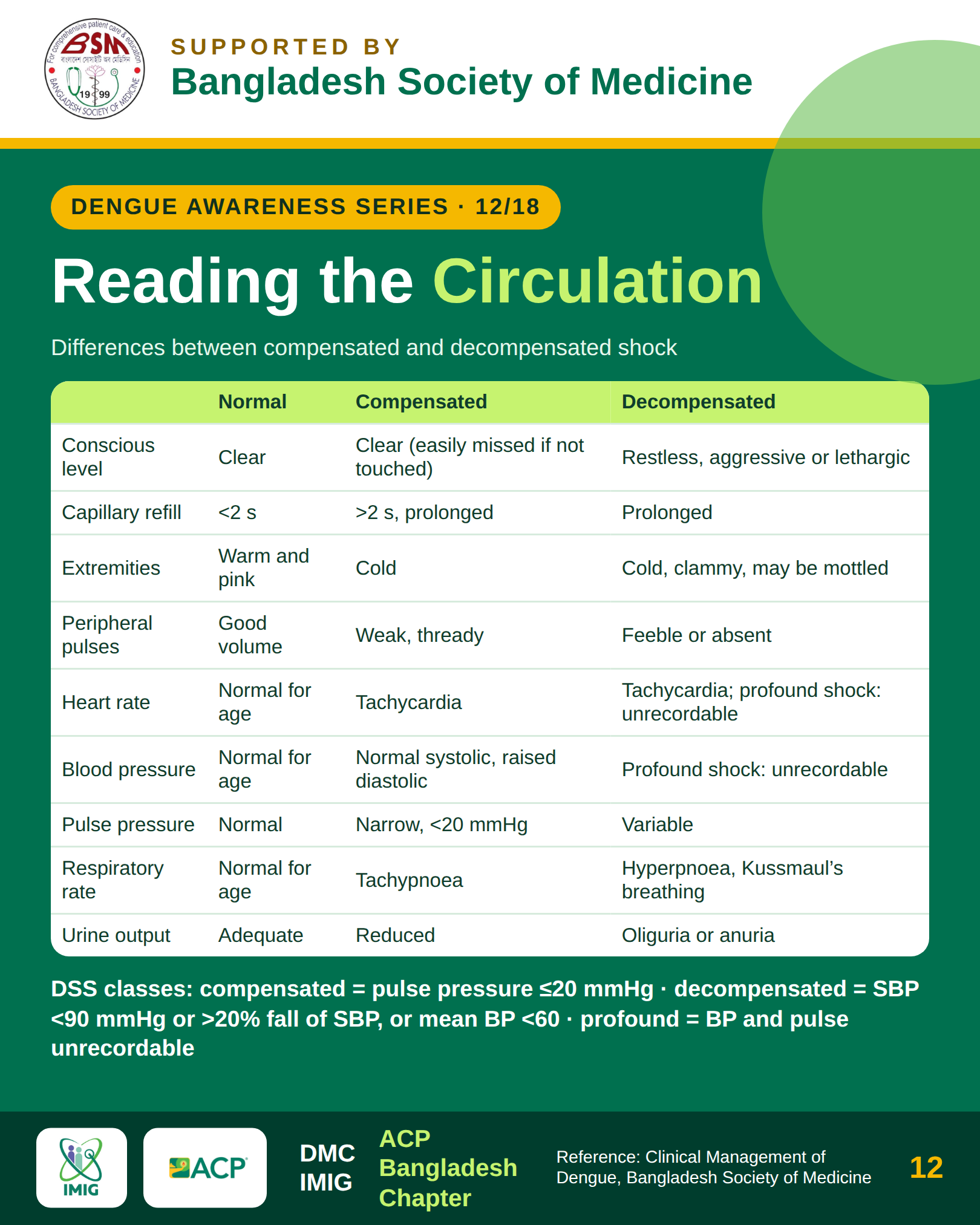
Recognizing Compensated vs. Decompensated Shock
- 1.Compensated Shock:
- Systolic blood pressure is maintained by intense peripheral vasoconstriction.
- Diastolic blood pressure rises, resulting in narrow pulse pressure (≤ 20 mmHg) (e.g., 100/85 mmHg).
- Tachycardia, delayed capillary refill time (> 2 seconds), and cool, pale peripheries.
- Urine output begins to decline (< 0.5 mL/kg/hr).
- 2.Decompensated Shock:
- Sympathetic vasoconstrictive compensatory mechanisms fail.
- Profound hypotension (systolic BP < 90 mmHg in adults or mean arterial pressure < 65 mmHg).
- Rapid, threadlike, or imperceptible peripheral pulses.
- Cold, clammy, cyanotic extremities with marked restlessness, confusion, or coma.
Section 5: Expanded Dengue Syndrome & Differential Diagnoses
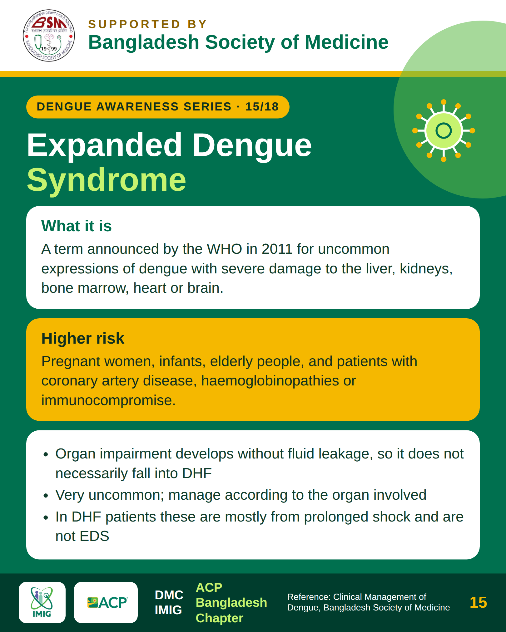
While plasma leakage remains the signature feature of DHF, Expanded Dengue Syndrome represents severe isolated or multi-organ failure that may develop even without marked plasma leakage or shock:
- Hepatic: Severe hepatitis, fulminant acute liver failure with massive transaminase elevation (AST/ALT > 1,000 U/L), prolonged prothrombin time, and hepatic encephalopathy.
- Neurological: Encephalitis, viral encephalopathy, acute disseminated encephalomyelitis (ADEM), transverse myelitis, and Guillain-Barre syndrome.
- Cardiovascular: Acute dengue myocarditis, arrhythmias, left ventricular systolic dysfunction, and sudden cardiogenic collapse.
- Renal: Acute kidney injury (AKI) resulting from prolonged hypoperfusion, rhabdomyolysis, or immune complex glomerulonephritis.
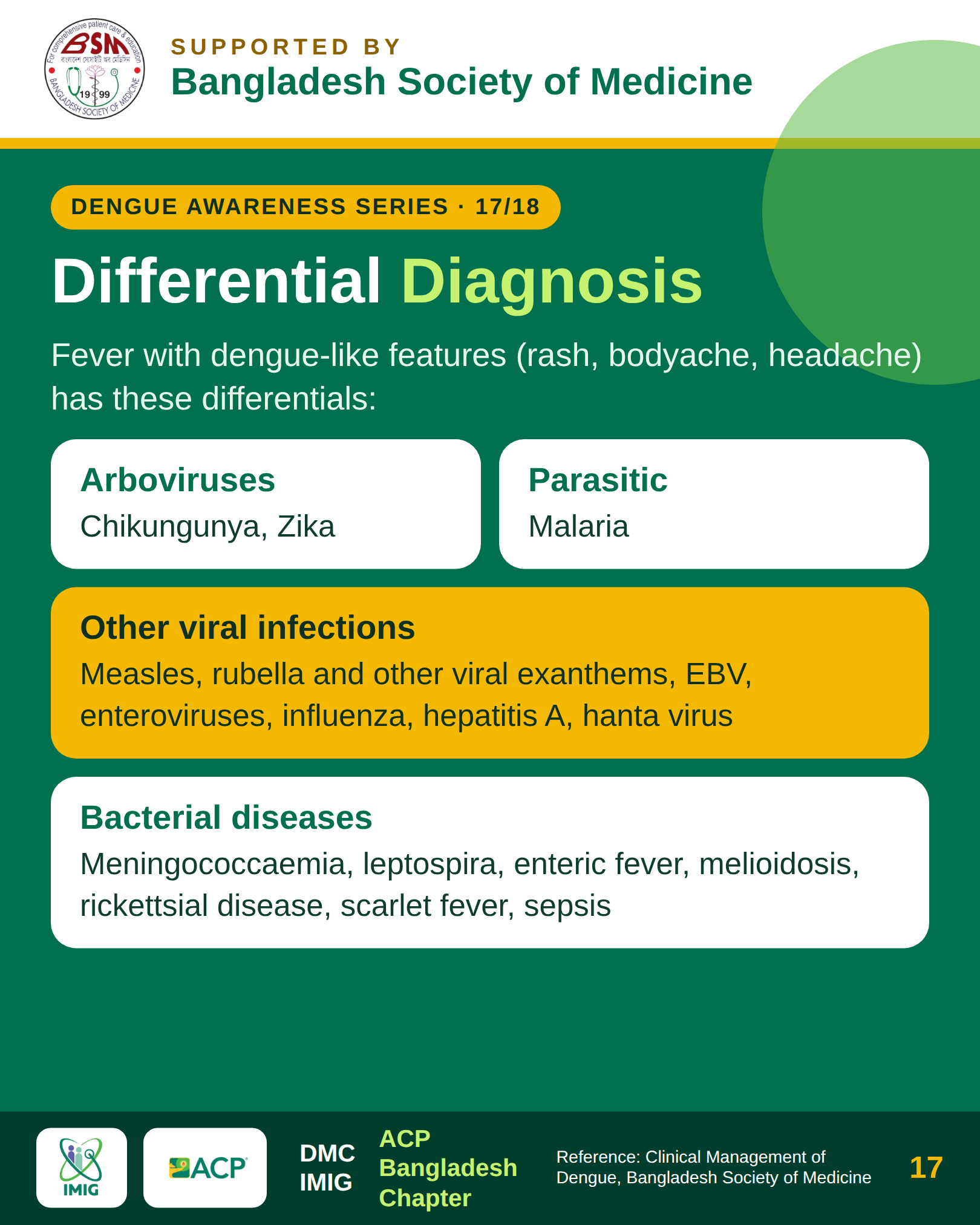
Differential Diagnosis of Acute Tropical Fevers
- Malaria: Rigors, periodicity of paroxysms, splenomegaly, diagnosed via thick/thin peripheral blood smears or rapid diagnostic tests (RDTs).
- Enteric (Typhoid) Fever: Step-ladder fever pattern, relative bradycardia (Faget's sign), coated tongue, leukopenia without profound thrombocytopenia, blood culture confirmation.
- Leptospirosis: Prominent conjunctival suffusion without purulence, severe calf tenderness, early jaundice, and azotemia.
- Chikungunya: Severe, debilitating, symmetric polyarthralgia and tenosynovitis with minimal plasma leakage.
Section 6: Complete Blood Count (CBC) & Hematocrit Dynamics
The Complete Blood Count (CBC) is the most indispensable test in dengue management, providing dynamic clues to disease trajectory when tracked longitudinally:
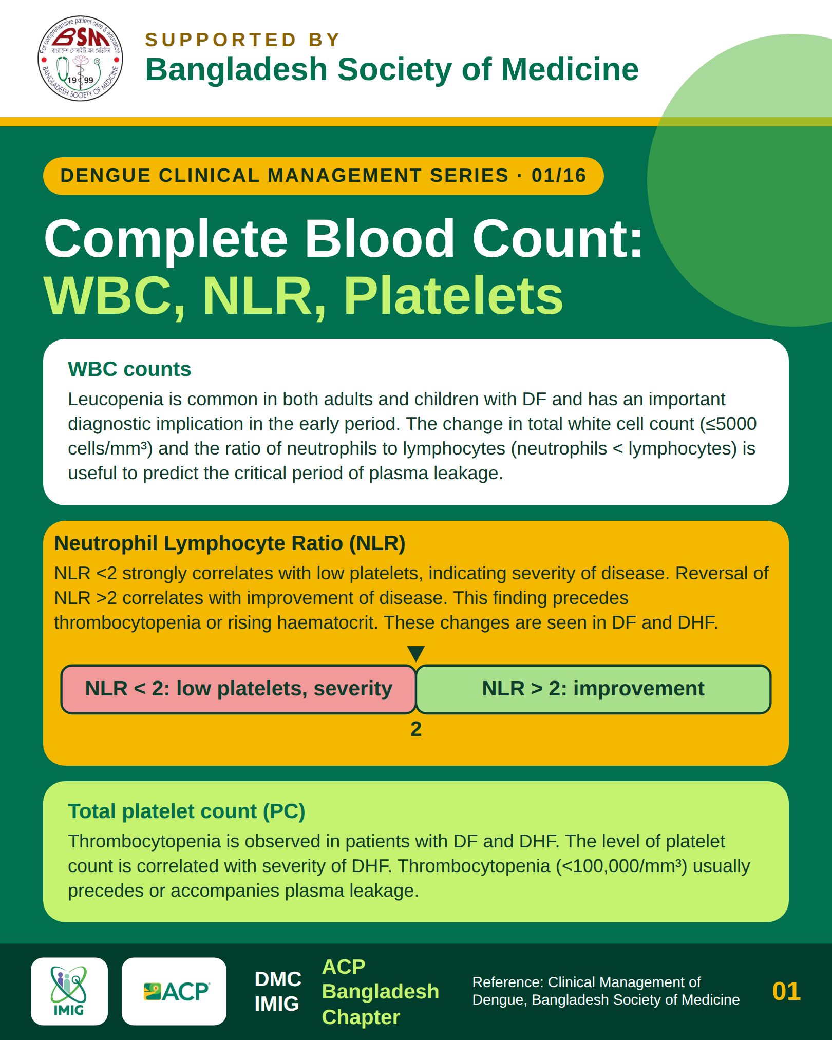
- White Blood Cell (WBC) Count: Leukopenia (WBC < 4,000/uL) is frequently the earliest hematological signal of dengue, appearing on Days 2 to 3. The nadir occurs just before the fever breaks. A rising WBC count during the critical phase warrants immediate investigation for secondary bacterial infection or occult major hemorrhage.
- Neutrophil-to-Lymphocyte Ratio (NLR): In early infection, neutropenia and relative lymphopenia occur. As the immune response matures, atypical reactive lymphocytes (Downey cells) surge, causing the NLR to drop significantly. A declining NLR often heralds the onset of the critical phase.
- Platelet Count: Progressive thrombocytopenia is universal in DHF. Platelets drop gradually during the febrile phase and reach their nadir between Days 4 and 6. A sharp drop (e.g., dropping from 120,000 to < 50,000/uL within 24 hours) is a major laboratory warning sign of impending plasma leakage.
Haematocrit (Hct): The Primary Bedside Guide
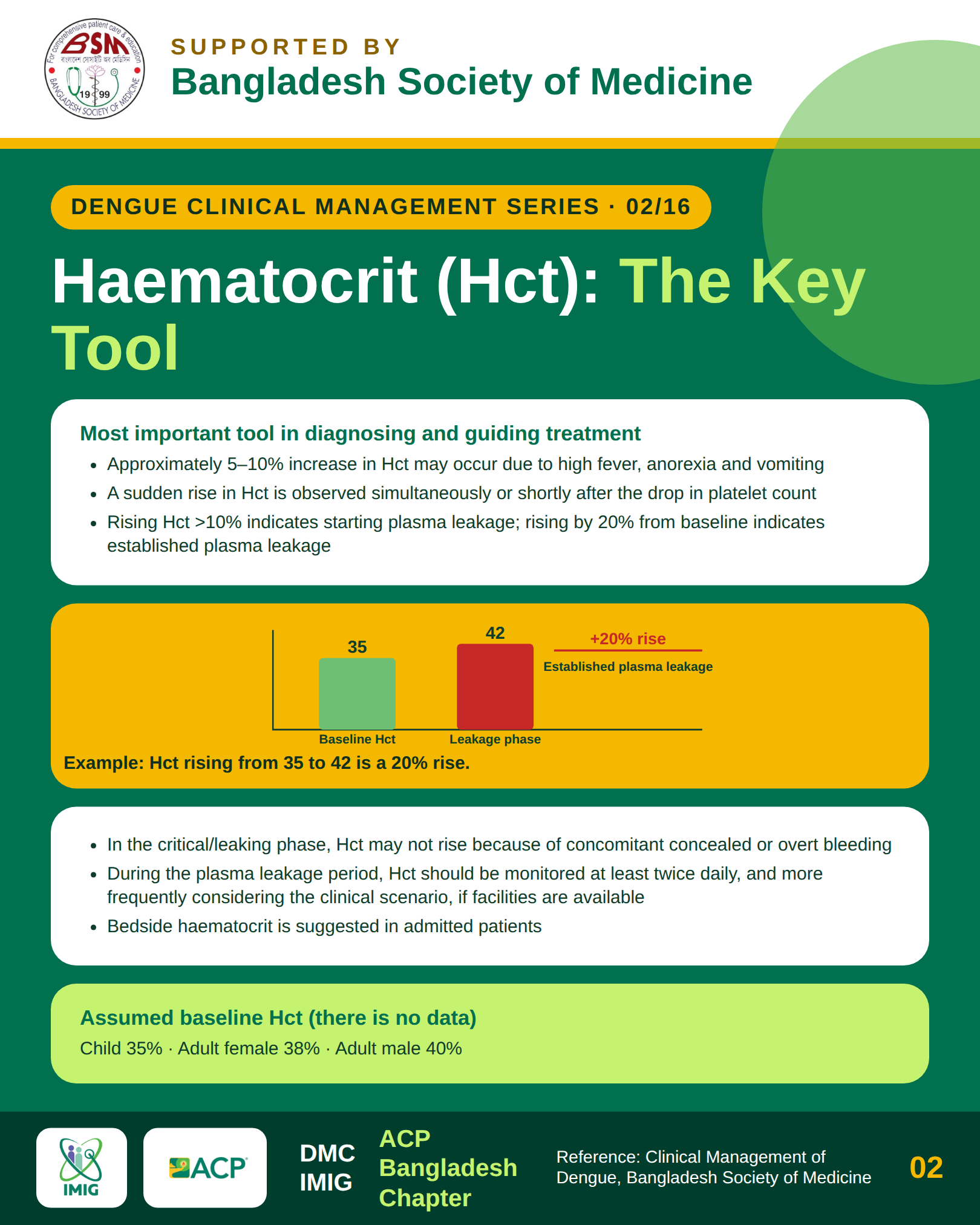
Hematocrit is the single most sensitive surrogate marker of intravascular volume and plasma leakage. Because fluid escapes into the extravascular third spaces while cellular elements remain inside the capillaries, hemoconcentration develops.
- Determining the Baseline Hematocrit: If a pre-illness or early febrile phase (Day 1) baseline is unavailable, population-specific age and gender estimates should be used (typically 40% to 45% in adult males; 36% to 40% in non-pregnant adult females).
- Formula for Percentage Rise in Hematocrit:
Percentage Hct Rise = [(Current Hct - Baseline Hct) / Baseline Hct] * 100 - 10% Rise Above Baseline: Early indicator that plasma leakage has begun. Warrants increased observation, reduction of physical exertion, and careful calculation of oral maintenance fluids.
- 20% Rise Above Baseline: Objective confirmation of significant plasma leakage and diagnostic criterion for DHF. Requires prompt initiation of titrated intravenous crystalloid therapy.
- Paradoxical Drop in Hct with Hemodynamic Instability: An unstable patient with dropping hematocrit indicates internal occult hemorrhage (most commonly upper gastrointestinal bleeding) rather than fluid stabilization. This mandates immediate blood transfusion rather than further crystalloid boluses.
Section 7: Diagnostic Windows & Laboratory Profiling
Selecting the correct diagnostic modality depends entirely on the day of illness:
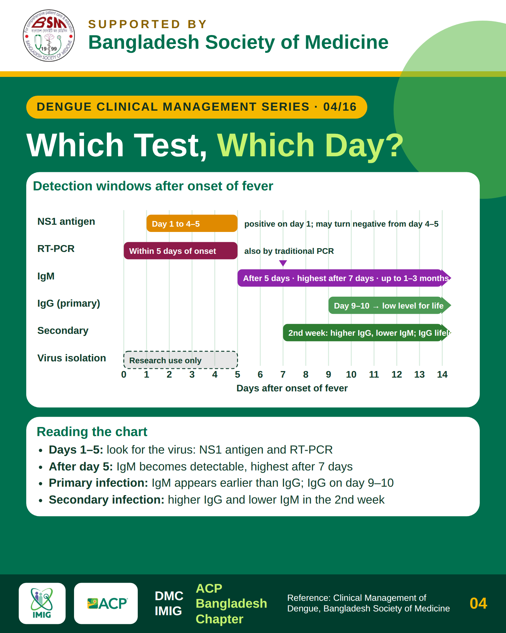
- Days 1 to 5 (Viremic / Early Febrile Phase):
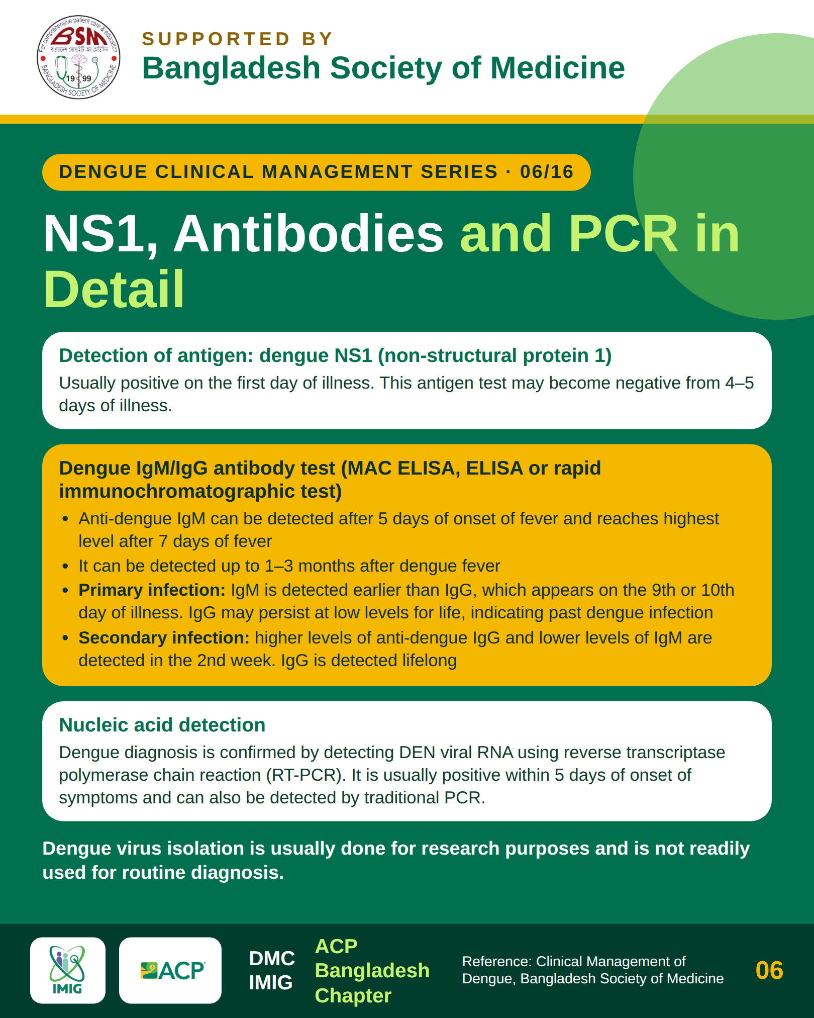
- Dengue NS1 Antigen: Highly sensitive and specific during the first 5 days. Circulating NS1 levels fall rapidly as antibody production begins.
- Reverse Transcription PCR (RT-PCR): Gold standard for confirming viral RNA, quantifying viral load, and identifying the specific serotype (DENV 1-4). Optimal on Days 1 to 4.
- Days 5 and Beyond (Serological / Defervescence Phase):
- Anti-Dengue IgM ELISA: Detectable starting around Day 4 to 5, peaking between Days 10 to 14, and persisting for 2 to 3 months. A positive IgM confirms recent dengue infection.
- Anti-Dengue IgG ELISA: In primary infection, IgG appears slowly after Day 10 in low titers. In secondary infection, IgG rises rapidly to very high levels within the first 48 hours of illness, often exceeding IgM titers.
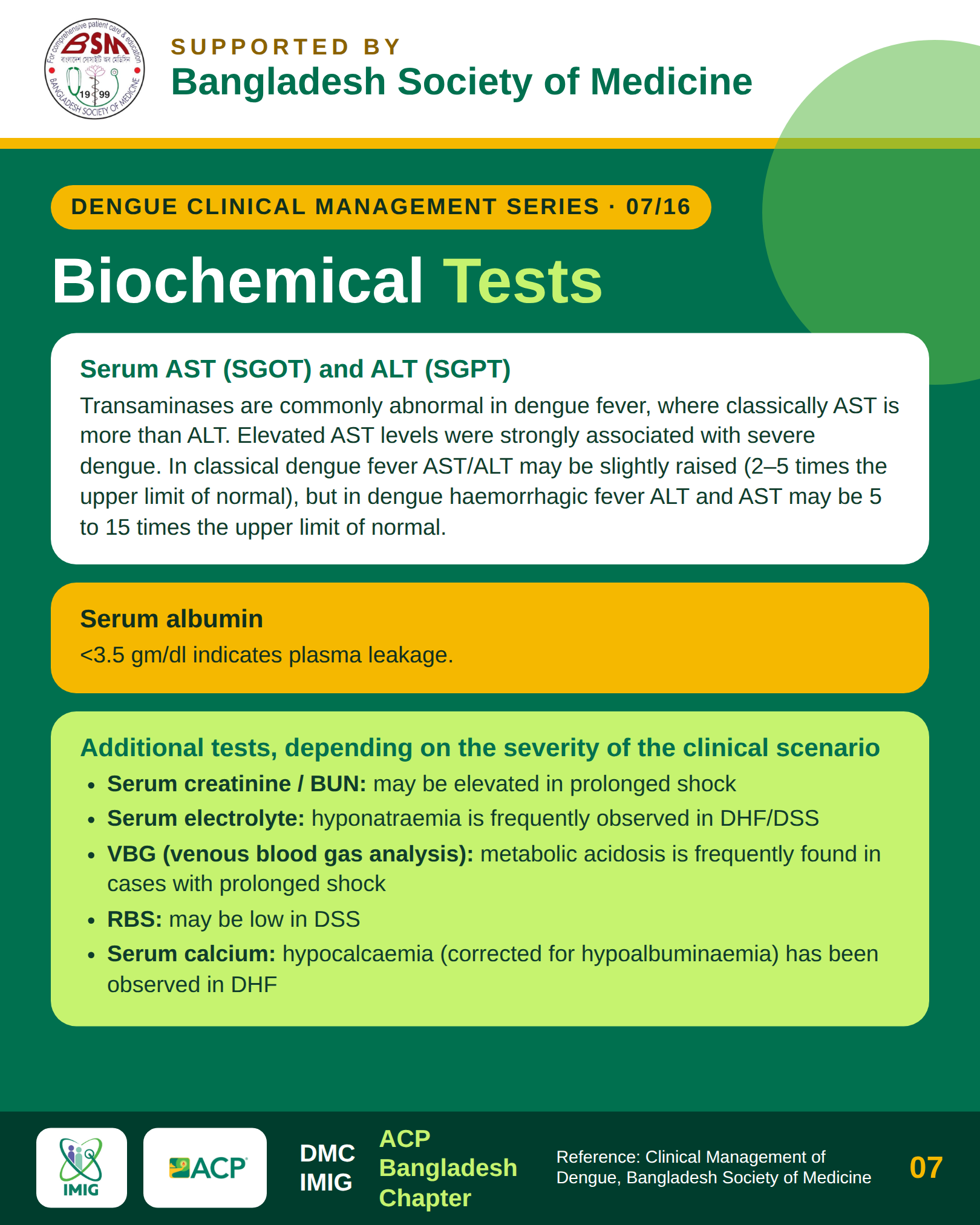
Biochemical & Coagulation Profiling
- Hepatic Transaminases: Aspartate aminotransferase (AST) and alanine aminotransferase (ALT) rise in the majority of symptomatic dengue patients. AST is characteristically higher than ALT in dengue, reflecting both direct hepatocyte injury and secondary release from damaged myocytes. Levels > 1,000 U/L indicate severe dengue-associated hepatitis or ischemic liver injury.
- Serum Albumin: Drops progressively during the critical leakage phase. Serum albumin < 3.0 g/dL strongly correlates with clinically significant pleural effusion and ascites.
- Electrolytes: Hyponatremia is the most common electrolyte disturbance in acute dengue, driven by inappropriate ADH secretion and gastrointestinal losses. Severe hyponatremia contributes to cerebral edema and encephalopathy.
- Coagulation Abnormalities: Mild prolongation of activated partial thromboplastin time (aPTT) and prothrombin time (PT) is frequent due to endothelial activation and transient depression of hepatic synthesis of clotting factors. Markedly prolonged PT/INR and aPTT alongside plunging fibrinogen (< 100 mg/dL) and elevated D-dimer signal consumptive coagulopathy (Disseminated Intravascular Coagulation, DIC).
Section 8: Neurological Complications & ICP Management
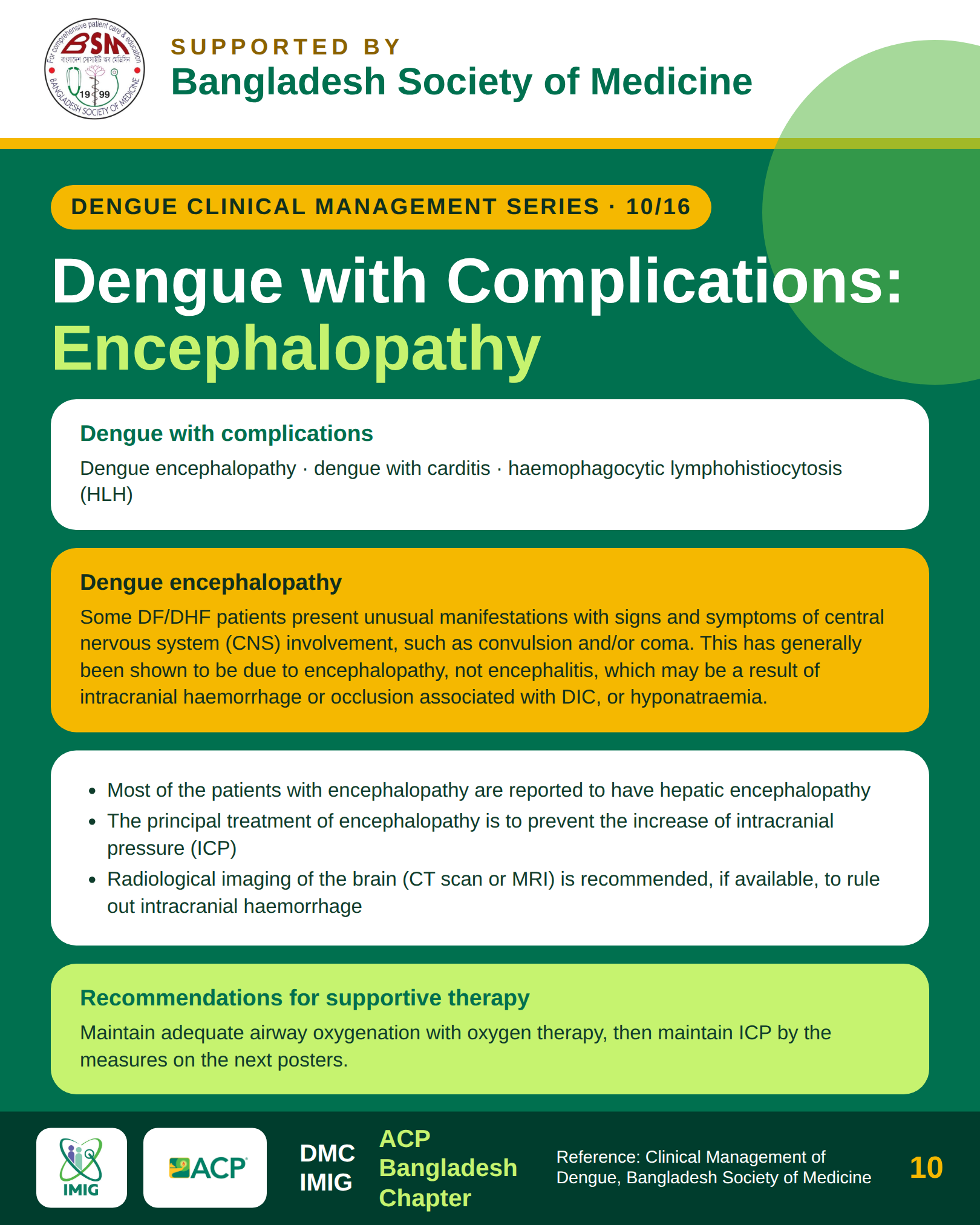
- Dengue Encephalopathy: Most common neurological manifestation, resulting from severe hepatic failure, prolonged hypoperfusion/shock, severe hyponatremia, or intracranial hemorrhage. CSF is characteristically normal without pleocytosis.
- Dengue Encephalitis: Caused by direct neurotropic invasion of the central nervous system by DENV. Confirmed by detection of DENV RNA or anti-dengue IgM in cerebrospinal fluid (CSF).
Management of Elevated Intracranial Pressure (ICP)
- 1.Elevate head of bed 30 degrees to promote cerebral venous drainage.
- 2.Maintain normothermia and prevent shivering.
- 3.Fluid restriction: Provide maintenance fluid titrated carefully to maintain cerebral perfusion pressure without aggravating cerebral edema.
- 4.Osmotic therapy: Intravenous 3% hypertonic saline (3 to 5 mL/kg over 15 to 30 minutes) or 20% mannitol (0.5 to 1.0 g/kg over 20 minutes) for acute neurological deterioration with signs of herniation.
- 5.Intubation and targeted mild mechanical hyperventilation (PaCO2 30 to 35 mmHg) as a temporizing emergency bridge.
Section 9: Secondary HLH & Critical Safety Red Lines
Secondary Hemophagocytic Lymphohistiocytosis (HLH) is an underrecognized, hyperinflammatory syndrome triggered by severe dengue, characterized by uncontrolled macrophage activation, hemophagocytosis, and overwhelming cytokine release.
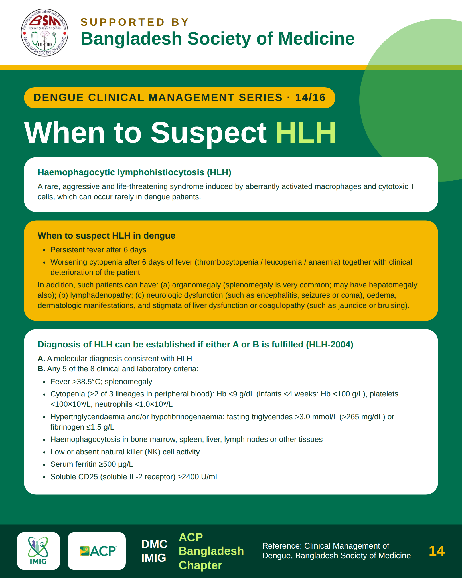
Diagnostic Red Flags (HLH-2004 Modified Criteria)
- Persistent high-grade fever lasting beyond Day 7 (failing to defervesce).
- Progressive bicytopenia or pancytopenia despite hemodynamic stability.
- Extreme Hyperferritinemia: Serum ferritin > 10,000 ng/mL is highly sensitive and specific for secondary HLH.
- Hypertriglyceridemia (fasting triglycerides ≥ 265 mg/dL) and hypofibrinogenemia (≤ 150 mg/dL).
- Splenomegaly and persistent transaminitis.
Treatment Protocol for Dengue-Induced HLH
- Pulse Intravenous Methylprednisolone: 500 mg to 1,000 mg IV daily for 3 consecutive days, followed by oral steroid taper.
- Intravenous Immunoglobulin (IVIG): 1 to 2 g/kg total dose administered over 2 to 5 days, particularly effective in refractory cases with profound thrombocytopenia and bleeding.
- Early multidisciplinary care involving hematology and intensive care specialists.
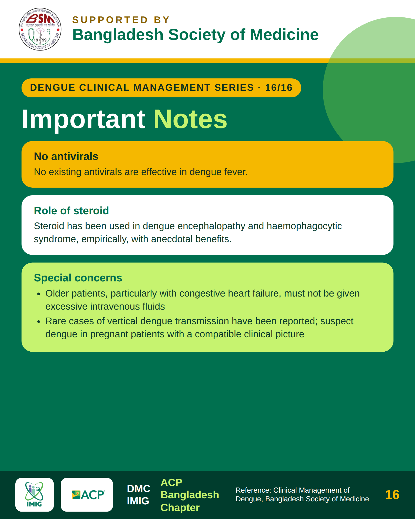
Critical Clinical Red Lines & Absolute Safety Rules
- 1.Never Administer NSAIDs or Aspirin: Ibuprofen, ketorolac, naproxen, and aspirin provoke severe gastric erosions and irreversible platelet inhibition, dramatically increasing life-threatening gastrointestinal hemorrhage. Only paracetamol (acetaminophen) at recommended doses (maximum 3 g/day in adults) should be utilized.
- 2.Never Give Prophylactic Platelet Transfusions: Low platelet counts alone (even < 20,000/uL) do not mandate transfusion in the absence of active major bleeding. Prophylactic transfusions do not prevent bleeding, fail to raise counts significantly due to rapid peripheral destruction, and increase risks of fluid overload, TRALI, and hospital length of stay.
- 3.Never Prescribe Routine Systemic Steroids in Uncomplicated Dengue: Corticosteroids provide no proven benefit in preventing plasma leakage or shock and increase the risk of secondary fungal and bacterial superinfections and hyperglycemia. They are reserved strictly for recognized immunologic complications like secondary HLH or refractory immune thrombocytopenia.
- 4.Enforce Fluid Safety Quotas: Intravenous fluid administration during the critical leakage phase must not exceed the calculated total quota (Maintenance + 5% Deficit). When the 48-hour critical leakage period ends, all intravenous fluids must be stopped immediately to avoid acute pulmonary edema during the convalescent reabsorption phase.
Explore the Complete Educational Series
- Complete 34-Infographic Dengue Awareness Gallery: Browse the entire collection of clinical slides in full resolution with interactive category filtering.